
Seventeenth century skull trepanation set
|
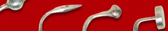
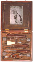
Silk and velvet dressing (5x20,5x18,5cm). Attached photograph of its prior owner. Four trephine crowns: steel; 2x7,2x2cm and 2x7,5x2cm Trephine handle: wood and steel; 6x13x2,5cm Rougine and lenticular knife: wood and steel; 2,5x17,5x2,5cm; Perforator: steel; 0,4x8,5x1,4cm Tweezer: steel; 4x13,5x4cm Double elevator: steel; 1,5x17x4cm Brush: ivory and bristle; 1x7x2,1cm Key: steel; 0,6x4x1,7cm Wood, ivory, bristle, steel, brass and cloth; 18th century |
It was mainly through Flemish painting of the 16th and 17th centuries that a scene of a skull trepanation could be visualized, with evidence to the position and expression of the patient, the position of the surgeon and his assistants, the technique and the instruments used. It was about the belief in the "Madness Stone" and the solution to this charlatanism was a skull trepanation. Berengario de Carpo (1518) made the first detailed description of the trepan. In the 17th century J. Scultetus in his "Armamentarium Chirurgicum" (1655), in the 18th century Heister in "A General System of Surgery" (1845), and in the next century Sir Charles Bell in "Illustrations of the great operations of surgery" (1821) and J. M. Bourgery and N. H. Jacob in "Traité Complet de l'Anatomie de l'Homme Comprenant la Médicine Opératoire" ("Complete Textbook of Human Anatomy Including Operating Medicine") (1837- 40), present images of a skull trepanation surgical scene, as well as the necessary surgical instruments which shapes and compositions are enhanced.
According to António de Almeida the chosen 17th century skull trepanation set, although without any manufacturer marks, has probably an English origin, considering the shape of the surgical instruments (gimlet handle and cylindrical trephine crowns).
In the period prior to the antisepsis/asepsis the instruments were continuously used in the patients without any concern with sterilization. If there was any instrument cleansing it was due to proper care and performance of the instrument itself. So, there was no obstacle to the inclusion of organic materials in the manufacture of such devices. The technological level and the scientific and technical demands of those times determined the use of ferrous and non-ferrous metals, which were then available.